New York Stem Cell Foundation (NYSCF) Research Institute collaborates with Google Research to identify new cellular characteristics of disease in skin cells from Parkinson’s patients.
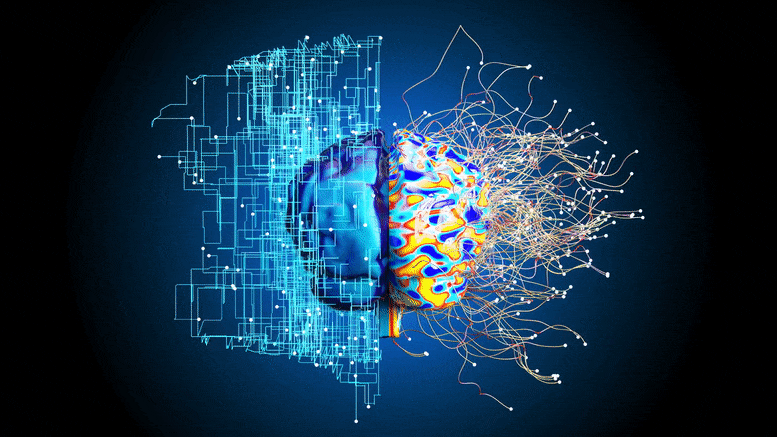
Copyright: scitechdaily.com – “Hidden Signatures of Parkinson’s Disease Uncovered by Artificial Intelligence and Robotics”
 A study published today (March 25, 2022) in Nature Communications unveils a new platform for discovering cellular signatures of disease that integrates robotic systems for studying patient cells with artificial intelligence methods for image analysis. Using their automated cell culture platform, scientists at the NYSCF Research Institute collaborated with Google Research to successfully identify new cellular hallmarks of Parkinson’s disease by creating and profiling over a million images of skin cells from a cohort of 91 patients and healthy controls.
“Traditional drug discovery isn’t working very well, particularly for complex diseases like Parkinson’s,” noted NYSCF CEO Susan L. Solomon, JD. “The robotic technology NYSCF has built allows us to generate vast amounts of data from large populations of patients, and discover new signatures of disease as an entirely new basis for discovering drugs that actually work.”
“This is an ideal demonstration of the power of artificial intelligence for disease research,” added Marc Berndl, Software Engineer at Google Research. “We have had a very productive collaboration with NYSCF, especially because their advanced robotic systems create reproducible data that can yield reliable insights.”
The study leveraged NYSCF’s vast repository of patient cells and state-of-the-art robotic system – The NYSCF Global Stem Cell Array® – to profile images of millions of cells from 91 Parkinson’s patients and healthy controls. Scientists used the Array® to isolate and expand skin cells called fibroblasts from skin punch biopsy samples, label different parts of these cells with a technique called Cell Painting, and create thousands of high-content optical microscopy images. The resulting images were fed into an unbiased, artificial intelligence–driven image analysis pipeline, identifying image features specific to patient cells that could be used to distinguish them from healthy controls.
“These artificial intelligence methods can determine what patient cells have in common that might not be otherwise observable,” said Samuel J. Yang, Research Scientist at Google Research. “What’s also important is that the algorithms are unbiased — they do not rely on any prior knowledge or preconceptions about Parkinson’s disease, so we can discover entirely new signatures of disease.”[…]
Thank you for reading this post, don't forget to subscribe to our AI NAVIGATOR!
Read more: www.scitechdaily.com


New York Stem Cell Foundation (NYSCF) Research Institute collaborates with Google Research to identify new cellular characteristics of disease in skin cells from Parkinson’s patients.
Copyright: scitechdaily.com – “Hidden Signatures of Parkinson’s Disease Uncovered by Artificial Intelligence and Robotics”
“Traditional drug discovery isn’t working very well, particularly for complex diseases like Parkinson’s,” noted NYSCF CEO Susan L. Solomon, JD. “The robotic technology NYSCF has built allows us to generate vast amounts of data from large populations of patients, and discover new signatures of disease as an entirely new basis for discovering drugs that actually work.”
“This is an ideal demonstration of the power of artificial intelligence for disease research,” added Marc Berndl, Software Engineer at Google Research. “We have had a very productive collaboration with NYSCF, especially because their advanced robotic systems create reproducible data that can yield reliable insights.”
Coupling Artificial Intelligence and Automation
The study leveraged NYSCF’s vast repository of patient cells and state-of-the-art robotic system – The NYSCF Global Stem Cell Array® – to profile images of millions of cells from 91 Parkinson’s patients and healthy controls. Scientists used the Array® to isolate and expand skin cells called fibroblasts from skin punch biopsy samples, label different parts of these cells with a technique called Cell Painting, and create thousands of high-content optical microscopy images. The resulting images were fed into an unbiased, artificial intelligence–driven image analysis pipeline, identifying image features specific to patient cells that could be used to distinguish them from healthy controls.
“These artificial intelligence methods can determine what patient cells have in common that might not be otherwise observable,” said Samuel J. Yang, Research Scientist at Google Research. “What’s also important is that the algorithms are unbiased — they do not rely on any prior knowledge or preconceptions about Parkinson’s disease, so we can discover entirely new signatures of disease.”[…]
Thank you for reading this post, don't forget to subscribe to our AI NAVIGATOR!
Read more: www.scitechdaily.com
Share this: